

Mucoid Pseudotumor Complicating a Pneumatocele that Developed as a Complication of Pulmonary Tuberculosis
Amit G. Kachalia, Erik Perez, Kinjal A. Kachalia, Jaspreet Kaur, Rahman Habibur
Med Sci Case Rep 2016; 3:48-51
DOI: 10.12659/MSCR.898696
Available online: 2016-06-03
Published: 2016-06-03
BACKGROUND:
Pneumatoceles usually develop as sequel of acute bacterial pneumonias and rarely as a complication of tuberculosis. Pseudotumors are usually inflammatory. We report the first case of a mucoid pseudotumor complicating a pneumatocele and one of the few reports of a pneumatocele that developed as a complication of pulmonary tuberculosis.
CASE REPORT:
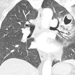
A 53-year-old female smoker with family history of lung cancer presented with chronic cough, weight loss, and fever. QuantiFERON-TB Gold was positive and a chest x-ray revealed a large homogeneous density in the left upper lobe. Computerized tomography of the chest showed pneumonia and a pneumatocele formation. Bronchoalveolar lavage grew acid-fast bacilli and Penicillium species; trans-bronchial biopsies showed nonspecific inflammation. The patient received treatment for Class 3 tuberculosis with symptomatic improvement. Later, she presented with cough and hemoptysis. Surveillance imaging showed increasing soft-tissue density and nodules in the area of previous consolidation, suggestive of a solid tumor. Positron emission tomography (PET) showed the affected area was minimally avid with central hypo-attenuation. Several weeks later, the patient returned with complete resolution of her symptoms after her protracted cough lead to expectoration of copious sputum and a large ball-like mucous formation. Repeat imaging showed clearing of the soft-tissue density and reappearance of pneumatocele. The patient has remained asymptomatic.
CONCLUSIONS:
Symptoms that relapse with radiological presentation would have warranted an extensive malignancy workup. This first case report of a mucoid pseudotumor complicating a pneumatocele case reminds us to include tuberculosis as one of the etiological organisms for pneumatoceles, and highlights the importance of continued surveillance, especially in smokers.
Keywords: Antitubercular Agents, Mycobacterium tuberculosis