

Spontaneous Pneumothorax in a Patient with Advanced Scleroderma: A Case Report
Viki Kumar, Keerat Rai Ahuja, Nfn Anila, Muhammad Aurangzeb, Theo Trandafirescu
Med Sci Case Rep 2017; 4:34-36
DOI: 10.12659/MSCR.902080
Available online: 2017-04-21
Published: 2017-04-21
BACKGROUND:
Spontaneous pneumothorax may be primary or secondary. There are many causes of secondary spontaneous pneumothorax. Spontaneous pneumothorax in association with scleroderma has been rarely reported.
CASE REPORT:
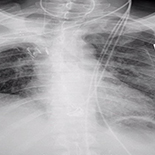
A 41-year-old man presented to the emergency department with sudden-onset dyspnea for 1 day. He denied having chest pain, palpitations, trauma, or other symptoms. Physical exam included mild tachycardia, tachypnea, Raynaud’s phenomenon, and sclerodermal skin changes on extremities, face, and trunk. Lab data included anti-topoisomerase >8 and antinuclear antibody (1: 2560). A chest x-ray revealed a large left pneumothorax with marked compression of the lung field and right-sided tracheal deviation. The patient’s past medical history included 1 year of severe progressive sclerosis, recurrent pneumothorax on right side status post chest tube placement, multiple bronchoscopies, right-sided video-assisted thoracoscopic surgery for persistent air leak, and muscle-sparing thoracotomy with right upper lobectomy. This time, the pneumothorax had recurred on the left side. A chest tube was inserted, resulting in lung expansion with symptom resolution. A post-chest tube CT scan chest showed severe paraseptal and centrilobular emphysematous changes with biapical bullous changes marked in the left apex. Within 3 days, the patient had another episode of dyspnea, with a chest x-ray showing recurrent left pneumothorax. Manipulating and changing the chest tube did not bring any improvement, so a blood patch pleurodesis was done. No recurrence was noted in the next 4 weeks.
CONCLUSIONS:
Physicians should consider pneumothorax as one of the potential complications in patients with chronic scleroderma with underlying pulmonary fibrosis and sub-pleural cysts.
Keywords: Pneumothorax, Scleroderma, Diffuse